視頻報道
-
椎管內(nèi)囊腫病例:藏在脊柱中的惡魔
CCTV-4《中華醫(yī)藥》
-

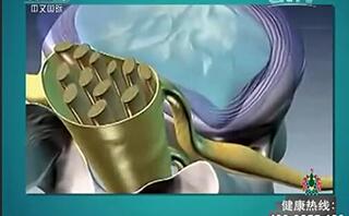
突出的腫瘤-腦膜瘤
CCTV10《走進(jìn)科學(xué)》
相關(guān)文章
Peritumoral edema shown by MRI predicts poor clinical outcome in glioblastoma
2016-12-15 17:51 作者:三博腦科醫(yī)院
Chen-Xing Wu1?, Guo-Shi Lin2?, Zhi-Xiong Lin1,2*, Jian-Dong Zhang2, Shui-Yuan Liu2 and Chang-Fu Zhou2
Abstract
Background: Magnetic resonance imaging (MRI) plays an irreplaceable role in the preoperative diagnosis of glioma, and its imaging features are the base of making treatment decisions in patients with glioma, but it is still controversial whether peritumoral edema shown by MRI from preoperative routine scans are associated with patient survival. The aim of this study was to assess the prognostic value of preoperative MRI features in patients with glioblastoma.
Methods: A retrospective review of 87 patients with newly diagnosed supratentorial glioblastoma was performed using medical records and MRI data from routine scans. The Kaplan-Meier method and COX proportional hazard model were applied to evaluate the prognostic impact on overall survival of pretreatment MRI features (including peritumoral edema, edema shape, necrosis, cyst, enhancement, tumor crosses midline, edema crosses midline, and tumor size).
Results: In addition to patient age, Karnofsky performance status (KPS) and postoperative chemoradiotherapy, peritumoral edema extent and necrosis on preoperative MRI were independent prognostic indicator for poor survival. Furthermore, patients with two unfavorable conditions (major edema and necrosis) had a shorter overall survival compared with the remainder.
Conclusions: Our data confirm that peritumoral edema extent and necrosis are helpful for predicting poor clinical outcome in glioblastoma. These features were easy to determine from routine MRI scans postoperatively and therefore could provide a certain instructive significance for clinical activities.
Keywords: Glioblastoma, Prognosis, Peritumoral edema, Necrosis, MRI
Background
Glioblastoma multiforme (GBM) is the most prevalent malignant brain tumor in adults and accounts for 17% of intracranial tumors. To date, effective durable treatments in patients with GBM are still lacking, exhibiting a poor prognosis with a median overall survival rate of 9.4 to 19.0 months despite advances in multimodal treatments that combine surgery, radiation therapy, and chemotherapy .Magnetic resonance imaging (MRI) technology is a common and noninvasive diagnostic modality and was previously found to take examination in central nervous system disease in clinical and especially in the preoperative diagnosis of glioma. Moreover, MRI is not only a powerful tool to visualize changes in morphological abnormalities but also a direct reflection of biochemical changes in the tumor and surrounding tissue. MRI can have great utility in the diagnosis, grading, and management of patients with GBM as many of the physical manifestations of the patho- logic processes in GBM can be visualized and quantified using MRI. Better taking account of the correlation be- tween preoperative tumor imaging features and survival is therefore useful to clinic.
For patients with GBM, clinical data-including age, perioperative Karnofsky performance status (KPS), pathological molecular markers, tumor resection, adjuvant radiochemotherapy, and tumor imaging features (including necrosis and edema) have been found to correlate with survival. Moreover, a clear relationship among survival pattern with peritumoral edema and necrosis in GBM has not been established. Some tumor imaging features of preoperative MRI from conventional scans, such as peritumoral edema (PTE) extent, necrosis, enhancement, and the size of cyst,were considered to be correlated with patient survival.However, several reports showed that these features, such as PTE and necrosis, were not independent values of survival in patients with glioblastoma .
These controversial results therefore suggest that there remains a need to further evaluate whether PTE and necrosis on MRI are associated with patient survival because such data from routine imaging play an irreplaceable role during preoperative diagnosis and now are the kernel base of making treatment decisions in patients with glioma, clearly recognizing the relationship among them has a certain instructive significance for clinical practice. Here, the tumor imaging features (including PTE, edema shape, necrosis, cyst, enhancement, tumor crosses midline, edema crosses midline, and tumor size) from preoperative routine MRI scans were assessed, and the aim of our study was to examine whether these characters are more valuable prognostic markers in patients with primary glioblastoma.
Methods
Study samples
Clinical and preoperative MRI data of 87 patients treated with resection of newly diagnosed supratentorial GBM at Beijing Sanbo Brain Hospital of Capital Medical University between April 2009 and March 2013 were
 京公網(wǎng)安備 11010802035500號